
Development of a Fluorescent Imaging Probe Specific for Neutrophils
Travis Riedel, PhD — Published: February, 28, 2013
This reagent is a Cyanine7-conjugated, PEG-modified hexapeptide that specifically binds the formyl peptide receptor (FPR) of neutrophils. After tail vein injection of the agent, sites of inflammation where neutrophils have been recruited and activated can be imaged in vivo by near-infrared (NIR) fluorescence. This Neutrophil-Specific, NIR Fluorescent Imaging Agent is now available exclusively from Kerafast.
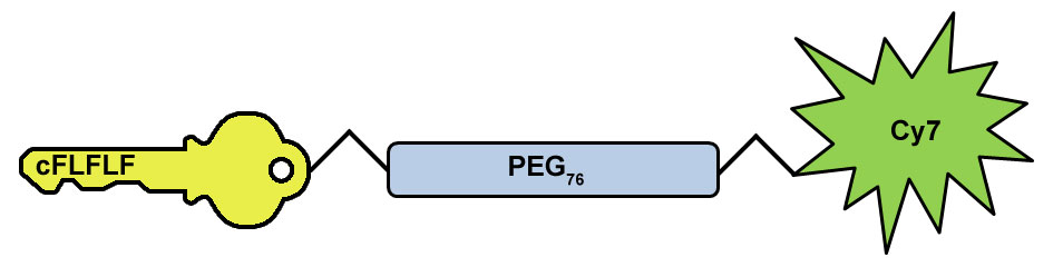
Diagram of Neutrophil-Specific, NIR Imaging Agent. Probe consists of cFLFLF FPR recognition peptide (gold key) and Cy7 NIR fluorophore (green starburst) connected via a PEG-76 hydrophilic linker (blue rectangle).
Over the past several years, real time in vivo fluorescence imaging of small animals has become a powerful tool for monitoring molecular events and biochemical processes in live animals. Dongfeng Pan’s laboratory at the University of Virginia specializes in the development of novel molecular imaging agents and therapeutics. In 2010, Dr. Pan published the synthesis of a Cyanine 7 (Cy7) labeled neutrophil-specific agent useful for near infrared fluorescence (NIR) imaging of live animals. The design centered around the cinnamoyl-F(D)LF(D)LF (cFLFLF) hexapeptide which has been shown to exhibit high affinity to the formyl peptide receptors (FPR) of neutrophils. This neutrophil-specific peptide was joined to Cy7, which, compared to other fluorophores, provides better photon signal penetration through tissue because of the minimal tissue absorbance in the NIR region of 700-900 nm.
Originally, the lab conjugated the peptide directly to the Cy7 fluorophore; however, the resulting compound was highly hydrophobic and not amenable to applications in live animals. To address this, a PEG-76 linker was added between cFLFLF and Cy7. This addition enhanced the overall bioavailability as demonstrated in hydrophobicity studies which measured the partition coefficient in n-octanol and water.
They then tested the imaging agent using a mouse model of acute inflammation in which phorbol myristate acetate (PMA) was applied topically on to the left earlobes of FVB mice. As a control, the right earlobes were treated with a vehicle consisting of DMSO and acetone. Additionally, as a measure of the imaging agent’s specificity for neutrophils, a group of PMA treated mice were administered a blocking agent consisting of the cFLFLF peptide conjugated to the PEG-76 linker, but lacking Cy7 fluorophore. After 1 hour, the Cy7 containing agent was injected and imaging experiments were performed.
Results from their studies showed a significant increase in fluorescence intensity of the PMA challenged ear compared to the control. Furthermore, mice treated with the blocking agent prior to administration of the Neutrophil Imaging Agent showed minimal fluorescence, demonstrating the high specificity of the probe for neutrophils.
Learn more about Neutrophil-Specific, NIR Imaging Agent »
Tags:
Live animal imaging, Neutrophils, Cyanine dye, formyl peptide receptor, Neutrophil-Specific, NIR Imaging Agent
References:
- Xiao et al. Synthesis of the Cyanine 7 labeled neutrophil-specific agents fornoninvasive near infrared fluorescence imaging. Bioorg. Med. Chem. Lett. 20 (2010) 3515–3517.

