Researchers create novel model system of the enteric nervous system
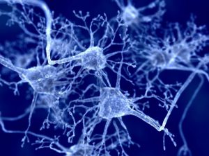
The gastrointestinal (GI) tract is innervated by the enteric nervous system (ENS) located within the GI tract, as well as through connections to the central nervous system (CNS). The ENS and CNS act in concert to control the homeostatic functions of the GI tract, such as digestion, secretion and motility. The constant, bi-directional communication between the two branches of the nervous system is known as the brain-gut axis. Interestingly, the ENS is capable of controlling GI functions independently of the CNS, and dysfunction of the ENS is associated with intestinal disorders such as inflammatory bowel disease and Hirschsprung’s disease.
A human ENS model system did not currently exist, hampering research in this area. Given the importance of the ENS in regulating GI function and disease, Dr. James Wells at Cincinnati Children’s Hospital and collaborators sought to develop a system that mimicked human intestinal tissue with a functional ENS. Their findings were recently published in Nature Medicine.
 Growing functional intestinal organoids
The first step in developing the human ENS model system involved growing human intestinal organoids in vitro. Organoids are 3D tissue cultures that recapitulate the organ of interest in structure and function. Wells and his collaborators previously published their work on growing human intestinal organoids (HIO) by differentiating human embryonic stem cells and induced pluripotent stem cells. These HIOs displayed the distinctive components of intestinal tissue (crypts, villi, intestinal stem cells, and smooth muscle), but lacked an ENS.
The next step was to incorporate an ENS into the intestinal organoids. Vagal neural crest cells (NCCs), the precursor to the ENS, were grown and aggregated with spheroids, tissues that had not yet differentiated into intestinal organoids. The aggregates were then grown in 3D culture conditions to create intestinal organoids with an immature ENS. HIOs and the immature ENS were then implanted into mice and monitored. The implanted HIOs + ENS matured and developed neurons and glia similar to the human small intestine.
The HIOs + ENS were found to be structurally similar to human small intestine, but were they functional? The researchers tested functionality by stimulating the HIOs + ENS and measuring for normal intestinal responses. The HIOs + ENS were able to release calcium, an important second messenger, in response to stimuli. Additionally, the HIOs + ENS could initiate wave contractions when stimulated, mimicking intestinal motility.
What does this mean for human research?
Rodent models are commonly used in gastrointestinal research; however, rodents differ from humans in gastrointestinal development, physiology and intestinal diseases, making them less than ideal models for human research. The model system created by Wells and collaborators provides the first human intestinal and ENS model system that is both physiologically and functionally similar to human intestine, allowing for more accurate study of human physiology and disease.
Related information
For more information on the GI tract, check out our previous blog post, How Well Do You Know the GI Tract? You also may be interested in one of our related reagents:
- Neuro-Dissociate Medium from Western Australian Neuroscience Research Institute
- Beta III Tubulin, Neuronal Antibody from University of Virginia
- Tau Antibody from University of Virginia
- Tissue Preservation Chamber from Southwood Scientific
- Mouse Cortico-Hippocampal Neurons from East Carolina University
- Rat Cerebellar Granule Cell Line (Cb-E1A) from University of Rochester

