Visualizing the Rise of Colon Cancer
A new research technique helps visualize a previously invisible process, showing how colon cancer cells grow and spread. Researchers at Duke Cancer Institute have used a modeling system in mice to observe how stem cell mutations arise in the colon. These mutations eventually take over and become malignant, leading to cancer. Their research was published in Nature Communications.
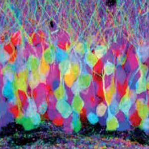
Brainbow Antibodies from University of Michigan.
The Technique
The researchers, Joshua Snyder, PhD and colleagues, have created a molecular dying technique named Crainbow (cancer rainbow) and applied it to colon cancer research. They are using the dying technique to tag common colon cancer mutations in the stem cells of a tumor to create a fluorescent barcode. These fluorescent stem cells can be tracked in the mouse to reveal the pre-cancerous cell activities.
In the study, the researchers cited our TagRFP Antibody from the Brainbow Antibody collection developed at the University of Michigan. According to the paper, the Crainbow modeling technique builds upon the original Brainbow method by combining it with functional genomics screening in one platform.
The Results
This technique has allowed for the researchers to discover the key differences in common intestinal habitats which grow malignant cancer cells. The difference happens at a critical period for newborns.
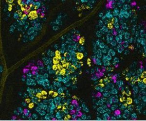
High magnification image of fluorescent intestinal stem cells. Each fluorescent color is used as a barcode to visualize colon cancer. Credit: Duke Health.
During this period, newborns are more susceptible to the negative effects of mutations in intestinal stem cells. If mutations occur, large fields of premalignant cells can grow throughout the intestine – known as field cancerization – without being detected by current screening protocols. These cells often remain harmless, unless exposed to the correct conditions in adulthood where they can turn cancerous. Therefore, field cancerization greatly increases the risk of cancer.
According to Snyder, “Field cancerization has been suggested to be the defining event that initiates the process of cancer growth, including cancers of the breast, skin and lung. Our technique allows us to model how premalignant cells compete and expand within a field by simple fluorescent imaging, potentially leading to earlier diagnosis and treatment.”
Moving Forward
While this study looked at colon cancer, additional studies are now looking at using the fluorescent barcoding technique to study breast cancer. For future breast cancer studies, the goal is to learn more about when ductal carcinoma in situ, a pre-cancerous condition, is driven by malignant or benign tumors.
Related Research
Do you work in this area of research? If so, you may be interested in viewing our other reagents for cancer research. We offer a variety of reagents closely related to our Brainbow Antibodies and colon cancer research, including:
- TagRFP [6A11f] Antibody from Fred Hutchinson Cancer Research Center
- tdTomato [16D7 Antibody] from The Scripps Research Institute
- mCherry [16D7] Antibody from The Scripps Research Institute
- mCherry [8C5.5] Antibody from University of Massachusetts Medical School
- GFP Antibody from The Scripps Research Institute
- MC-38 Cell Line from the National Cancer Institute/NIH

